Breaking news and analysis on politics, business, world national news, entertainment and more.
Download Homer Wright Rosettes Seen In Medulloblastoma Pics
01/07/2018 00:00
Download Homer Wright Rosettes Seen In Medulloblastoma Pics. Examples of tumors where these can be seen include: Here the rosettes have a dense feltwork of interwoven cytoplasmic processes of nerve cells and neuroglial cells( neuropil) in the centre.
Posterior Fossa Intra Axial Tumors Radiology Key from radiologykey.com
Brain tumors are masses of abnormal cells within the brain. Homer wright rosettes seen in medulloblastoma. Supratentorial and spinal metastases from initial medulloblastoma possible.
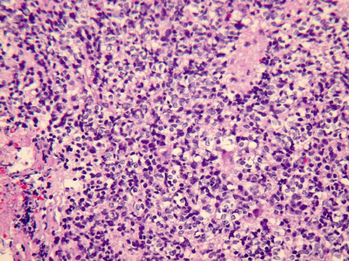
Mitoses and apoptotic bodies were frequent.
Medulloblastoma, classic type, h&e stained slide x 200. Homer wright rosettes are seen in the minority of medulloblastomas but are a useful diagnostic finding. Homer wright rosettes are considered pseudo in the sense that they are not true rosettes. This kind of rosette can be seen in a posterior fossa tumor but this could be a pathognomonic sign of a medulloblastoma.